
Endoscopic Ultrasound
Endoscopic Ultrasound
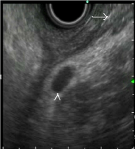
An endoscopic ultrasound scan uses an endoscope with an ultrasound probe attached to create detailed pictures of internal organs and structures.An endoscopic ultrasound scan (known as EUS) combines two types of test – endoscopy and ultrasound. The doctor uses an endoscope with an ultrasound probe attached to look inside your gut (gastrointestinal tract). By putting the endoscope into the upper part of the gut, EUS can create pictures of the surrounding structures, not just inside the gut. The ultrasound probe is used to create detailed pictures of the body, including the lungs, pancreas, liver, gallbladder and stomach.EUS can also look at other structures lower down in the body by inserting the endoscope through the rectum into the lower part of the gastrointestinal tract.The tip of the endoscope contains a light and a tiny video camera so the operator can see inside your gut. In an endoscopic ultrasound scan the endoscope also contains an ultrasound probe.
The procedure and preparation are similar to upper gastrointestinal endoscopy or lower gastrointestinal colonoscopy.The endoscope also has a ‘side channel’ down which instruments can pass. These can be manipulated by the doctor to take a small sample (biopsy) by using a thin ‘grabbing’ instrument or a fine needle which is passed down a side channel.
About
Dr. Neelam Mohan is one of the few women doctors in India who has balanced the various pillars of medical profession and is appreciated as an astute clinician/ healer, bright teacher, researcher, efficient leader/ administrator and for her contribution in social work. She has to her credit many achievements that has put the country on the global medical map.
Services
Copyright 2019 Drneelammohan.com. All rights reserved.